Otolaryngology: Applied Basic Sciences for YO-IFOS
.png/:/rs=h:100,cg:true,m)

G-protein coupled receptor (GCPR), G-protein coupled cascade (Golf-cascade), cyclic adenosine monophosphate (cAMP), adenylate cyclase type III (ACIII)

Olfactory epithelium comprises three cell types
1. Olfactory neurons: bipolar neurons (primary sensory neuron)
- Dendrites (peripheral processes) extend to the epithelial surface where they expand into an olfactory knob with cilia, which contain the molecular receptor sites.
- Central processes transmit sensation: these assemble into twenty or more bundles (filaments) which pass through the cribriform plate of the ethmoid bone to synapse on the secondary sensory neurons in the olfactory bulb.
2. Sustentacular cells (supporting cells) are amongst the sensory cells and are like glial cells.
3. Basal cells lie on the basement membrane and are the source of new receptor cells. Cells are replaced approximately every 60 days. This is the only area of the central nervous system where cells are continuously regenerated throughout life.
Olfactory Bulb

- The olfactory bulb is the rostral enlargement of the olfactory tract. The olfactory bulbs and tracts are parts of the brain that evaginated from the telencephalon in early development. The olfactory bulb contains the cell bodies of the secondary sensory neurons involved in relaying olfaction to the brain. It contains:
- Spherical structures called glomeruli: site of contact between the primary olfactory neurons, and mitral cells and tufted cells of the secondary olfactory neurons. Periglomerular interneurons interact between glomeruli.
- There is a high incidence of convergence within the glomerular layer as well as input from the central nervous system. Neural convergence is the process by which neural impulses from multiple neurons impact a single neuron or neuron group.
- The tufted and mitral cells are functionally similar. Together they represent the afferent neurons from the olfactory bulb to the CNS.
1. Mitral cells: the axons of primary sensory neurons and interneurons contact the mitral cell dendrites in the glomerular layer. The mitral cell axons extend collateral fibres to the anterior olfactory nucleus and then mainly project to the lateral (primary) olfactory area.
2. Tufted cells: the axons of primary olfactory neurons synapse with tufted cell dendrites in the glomerular layer. The tufted cell axons then project to the anterior olfactory nucleus and to the lateral and intermediate areas.
Starting at the cribriform plate, the bulb is arranged in five layers:
1. The most superficial layer: the nerve fibre layer (olfactory axons) contains the axons of primary olfactory neurons in the nasal mucosa. Primary afferent neurons are continually replaced throughout life.These synapse with secondary neurons in the olfactory bulb, which is a direct extension of the cortex, before synapsing in the thalamus. All other sensory neurons initially synapse in the thalamus.
2. The glomerular layer contains spherical glomeruli. Considerable convergence takes place between the axons of primary olfactory neurons and the dendrites of tufted and mitral cells. There is also input from interneurons and centrifugal efferents from the central nervous system.
3. The external plexiform layer contains mainly the bodies of tufted cells.
4. The mitral cell layer is a single layer of large mitral cell bodies.
5. The granule cell layer contains granule cells (inhibitory interneurons) and myelinated axons of secondary neurons.
After studying the diagram. Click on the image, below, to review the video and reinforce this concept.

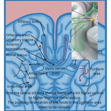
Olfactory tract and trigone
Postsynaptic fibres (of the olfactory bulb) form the olfactory tract and trigone. The trigone is an expansion of the olfactory tract just rostral to the anterior perforated substance of the brain. The fibres divide in front of the anterior perforated substance into lateral and medial olfactory striae to transmit impulses to olfactory areas for the conscious appreciation of smell.
The olfactory area
The limen insula, uncus, and entorhinal area are collectively called the pyriforrm (pear shaped) area.
The olfactory area consists of the cortices of the:
1. Uncus and
2. Entorhinal area (anterior part of the parahippocampal gyrus)
3. The limen insula (the point of junction between the cortex of the insula and the cortex of the frontal lobe), and
4. Part of the amygdaloid body (a nuclear complex located above the tip of the inferior horn of the lateral ventricle).
The primary olfactory area
.png/:/cr=t:0%25,l:0%25,w:100%25,h:100%25/rs=w:370,cg:true)
Olfactory Bulb Projections
Postsynaptic fibres (of the olfactory bulb) from the olfactory tract and trigone
Most of the axons from the olfactory tract pass via the lateral olfactory stria to the primary (lateral) olfactory area.
Some collateral branches of the axons of the secondary sensory neurons terminate in a small group of cells called the anterior olfactory nucleus. This is a collection of nerve cell bodies located along the olfactory tract. Postsynaptic fibres from this nucleus travel either with the central processes of the mitral and tufted cells or travel in the medial olfactory stria to enter the anterior commissure to reach the contralateral olfactory bulb. Their influence on the contralateral olfactory bulb is mainly inhibitory.
This serves to enhance the more active bulb and provide directional cues to the source of the olfactory stimulation. (See schematic representation of the olfactory bulbs and olfactory epithelium).
The olfactory system shares the entorhinal cortex with the limbic system, which has extensive connections with the septal area (formerly known as the medial olfactory area) of the frontal cortex and the hypothalamus with its autonomic centres.
From the primary olfactory areas, projections go to:
· The olfactory association area in the entorhinal cortex (Brodmann’s area 28)
· The hypothalamus
· The dorsal medial nucleus of the thalamus from which afferent neurons project to the orbitofrontal cortex for the conscious appreciation of smell.
Major Projections of the Olfactory Cortical Areas
Cells of the olfactory cortex have reciprocal connections with other regions in the olfactory cortex and outside the cortical areas. They contribute to fibres reaching the autonomic centres for visceral responses, such as salivation in response to pleasant cooking odours, nausea in response to unpleasant odours.
The principal pathways are:
- The medial forebrain bundle: information from all olfactory areas to the hypothalamus and brain stem reticular formation
- The stria medullaris thalami: olfactory stimuli from the various olfactory areas to the habenular nucleus (epithalamus)
- The stria terminalis: information from the amygdala to the anterior hypothalamus and the preoptic are
- The dorsal longitudinal fasciculus: information from the hypothalamus to the brain stem and spinal cord
The habenular nucleus and the hypothalamus project to the brain stem reticular formation and the cranial nerve nuclei, which are responsible for visceral responses. An example of this are superior and inferior salivatory nuclei and the dorsal vagal nucleus, which accelerate intestinal peristalsis and increase gastric secretion.

References
1. Kang N, Koo J. Olfactory receptors i n non-chemosensory tissues. BMB reports. 2012 Nov;45(11):612.
2. Morosanu CO, Humphreys C, Egerton S, Tierney CM. Woodruff's plexus-arterial or venous? Surg Radiol Anat. 2022 Jan;44(1):169-181. doi: 10.1007/s00276-021-02852-0. Epub 2021 Oct 29. PMID: 34714375.
3. Kamel, R., 2018. Understanding the complex anatomy of the middle turbinate via educational origami.
4. Al-Saigh NN, Harb AA, Abdalla S. Receptors Involved in COVID-19-Related Anosmia: An Update on the Pathophysiology and the Mechanistic Aspects. International Journal of Molecular Sciences. 2024 Aug 5;25(15):8527.
5. Nag AK, Saltagi AK, Saltagi MZ, Wu AW, Higgins TS, Knisely A, Ting JY, Illing EA. Management of post-infectious anosmia and hyposmia: a systematic review. Annals of Otology, Rhinology & Laryngology. 2023 Jul;132(7):806-17
